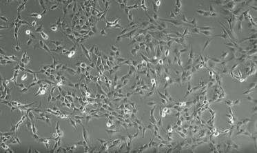
In 2018, we presented the results of our self-made animal serum-free medium (HELENE Medium) at the Cambridge International Stem Cell Symposium. The first point is that more cells can be cultured with HELENE Medium than with other commercially available media in the same period of time. The second point is that the size of the mesenchymal stem cells cultured with HELENE Medium is the smallest. The academic evidence suggests that the smaller the stem cells are, the more unlikely they are to differentiate into fibroblasts.
The above two points together confirm that the fat tissue taken from behind the ear does contain MSCs. The amount of fat collected is relatively low compared to that of the thigh or stomach, but with our own laboratory technology and HELENE Medium, it is possible to culture up to 2.25 billion MSCs in one month.
In order to assure the quality of the stem cells we provide to our patients, we have outsourced stem cell sample testing that is manufactured using the same process as our quality certificate. The testing complies with Good Manufacturing Practice (GMP) and Good Laboratory Practice (GLP) for regenerative medicine, and includes three independently designed experiments. We also provide cells to a third-party reagent company after conducting the same tests in our own laboratory, and we have verified the quality of the stem cells to be provided to patients in a fair manner.
Adipose tissue from three sample donors was cultured in our lab and sent for testing. The samples were taken from behind the ear, and the tissue was separated and broken down using special techniques and dedicated enzymes. The cells were made into single cells, cultured for one month, and those that reached the specified number were separated by MACS, and after the cell count was verified by cell counter, they were provided to a third-party reagent company, Takara Bio Inc.

| Test purpose | Test Title | Samples | Result |
|---|---|---|---|
| Infection and Contamination |
Sterility Test (Japanese Pharmacopoeia Direct Method, Option: Method Suitability Test) |
QC_AL Lot:#13349 | Negative |
| Sterility Test (Japanese Pharmacopoeia Direct Method) |
QC_AL Lot:#13349 | Negative | |
| Mycoplasma Exclusion Test (Japanese Pharmacopoeia Reference Information, PCR Method (with Confirmation of 7 Species Inhibition)) |
QC_AL Lot:#13349 | Negative | |
| Endotoxin Test (Japanese Pharmacopoeia Kinetic Turbidimetric Method, Option: Interfering Factors Test) |
QC_AL Lot:#13349 | Negative | |
| Endotoxin Test (Japanese Pharmacopoeia Kinetic Turbidimetric Method) |
QC_AL Lot:#13349 | Below detection accuracy | |
| Tumorigenesis | Colony Formation Test | QC_AL Lot:#13349 | Negative |
| Stem Cell Confirmation |
FCM Test (CD45- CD105+) |
QC_AL Lot:#13349 | CD45- 92.1% CD105+ 100% |
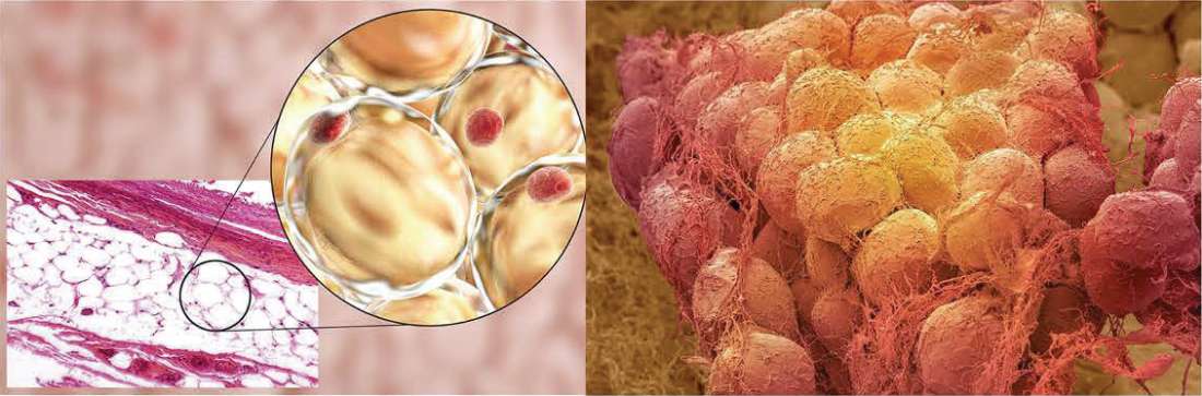
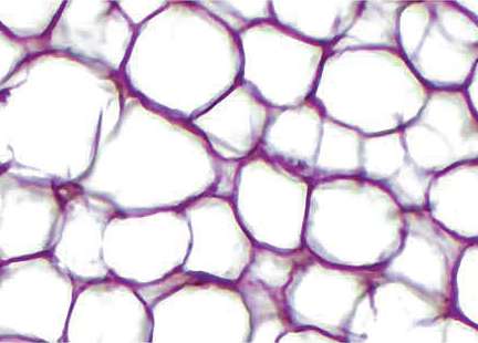
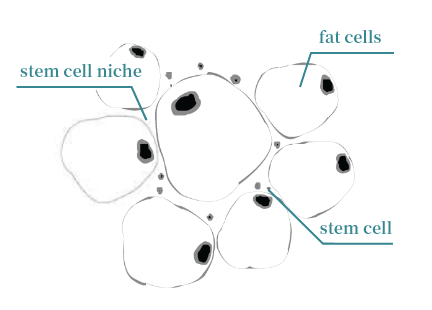
Below is an image of adipose tissue, and a round bean-like object is adipose tissue. It hides in a fibrous net-like part called the stem cell niche.
In other words, stem cells are not present in lipid droplets but are present in the surrounding fibrous areas. Let's take a look at the difference between back of the ear and belly fat
| Fat collect from back of the ear | abdominal subcutaneous fat | |
| Appearance photo | 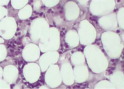 |
 |
| Image | 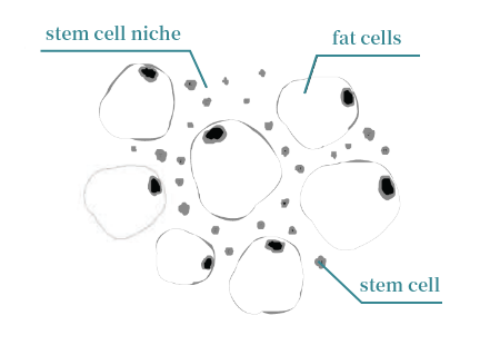 |
 |
| Pain | Less | Strong (Like hitting with the bruise) |
| Internal bleeding | Very rare | High possibility |
| Oppression | With bandage only (Recovery time : Short) |
Compress with cotton sheet (Recovery time : Long) |
Adipose-derived stem cells are not in yellow lipid droplets,
They are lurking around lipid droplets(= stem cell niche) called stroma. Stem cells are present in both abdominal fat and ear back fat, but the rate of stem cells is higher in "fat with less lipid droplets and more interstitial".
At Helene, we mainly collect sample behind-the-ear fat, which is less painful for patients.
However, for those who prefer abdominal fat, abdominal liposuction can also be used.
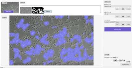
Stem cells cultured at HELENE Cell Center are measured in at least two stages to check the number and viability before they are offered to customers. The results are printed and lab-approved and will be provided to all customers in a scientific and responsible manner.
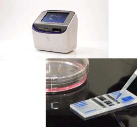
After obtaining the cell samples, staining is performed by adding a biological stain, 0.4% Typan blue solution, which is commonly used to differentiate living cells from dead cells. The staining method is a dye exclusion test, which takes advantage of the fact that the stain can penetrate into dead cells but is blocked by the cell membrane of living cells to distinguish them for the purpose of survival rate calculation. In the past, stained samples were observed through a microscope and counted manually, but we use an automated cell to automatically recognize the fluoroscopic response and complete the analysis. When counting manually, the user must not only adjust the focus manually, but also adjust the bright field light intensity between samples. The Countess II FL automated cell counter automatically adjusts the illumination and focus to obtain the best image quality for use.
After the quantity is measured using Countess II FL, our cell center will place the cell sample on a slide and obtain cell images with a ZEISS Axio Vert.A1 microscope. Using HELENE's self-developed AI program to read the images, the cell skeleton is observed at the confocal level, and the number of cells and cell viability are calculated by combining the focal length of the microscope, the cell type (MSC), and the cross-sectional area of the cell incubated flask. The precision of the two stages is close to 100%, achieving an artisan process that cannot be matched elsewhere, thus ensuring that HELENE brand therapeutic-graded stem cells are precisely tailored to customer needs and continuously optimized.
HELENE Clinic produces the certificate of stem cells for each treatment to every customer to show the responsibility of quality and transparency.
According to the paper [The safety of MSC therapy over the past 15 years: a meta-analysis], published in 2021, 62 studies were selected for analysis, including extracts from clinical trials published in the internationally renowned journal Lancet. Many of these trials have delivered more than 500 million cells, and some have delivered more than 1 billion cells, dispelling myths and confirming the technical strength of the HELENE Clinic to be in line with world-class research institutions.
| Author | Year | Location | Dose | |
|---|---|---|---|---|
| Hess | 2017 | USA | 1200×106 cells | 1,200 million MSCs |
| Bartunek | 2013 | Belgium | 600 -1200×106 cells | 600-1,200 million MSCs |
| Chanbers | 2017 | Australia | 1×10⁹ cells | 1,000 million MSCs / Lung disorder |
| Steinberg | 2017 | USA | 1×10⁹ cells | 1,000 million MSCs / Stroke |
| Igresias | 2021 | Mexico | 1×10⁹ cells | 1,000 million MSCs / Lung disorder |
| Matsuoka | 2024 | Japan | 1-2×10⁹ cells | 2,000 million MSCs / AntiAging |
We may not find clinical data where low cell counts were administered for specific diseases, suggesting that in some cases lower doses of MSC may be ineffective. As science progresses, treatment protocols and targets, sources and culture methods, and dosage differences of MSC will be continually improved and standardized. Until then, HELENE Clinic will keep refining the treatment approach, adapting different MSC and MSC-exosome volume transfusion base on the age, medical condition and disease of each patient to ensure the best possible stem cell treatment in complete safety.
[Quote] The safety of MSC therapy over the past 15 years: a meta-analysis.
https://doi.org/10.1186/s13287-021-02609-x
| Consultation hours | 10:00~19:00 |
|---|---|
| Closed days | Wednesday · Sunday |
| Address | 〒107-0062 5-9-15 Minami Aoyama, Minato-ku, Tokyo Aoyama OHMOTO Building 3F |
| Tel | 03-3400-2277 |