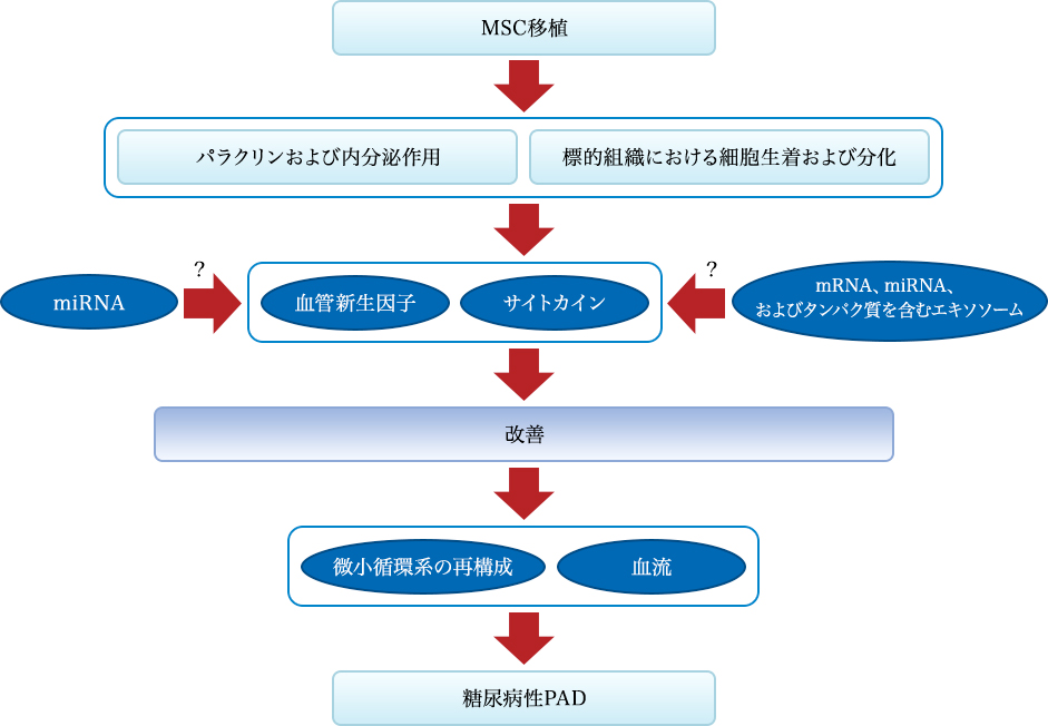
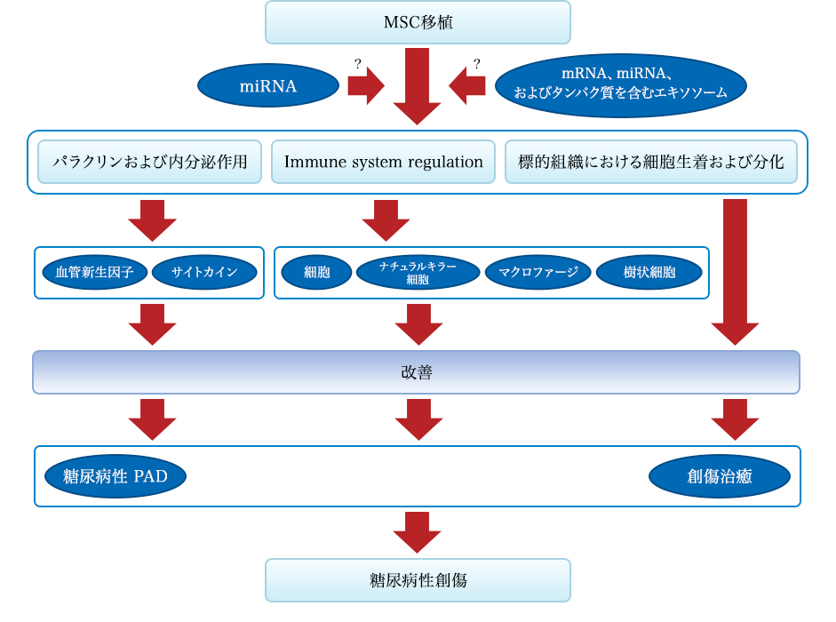
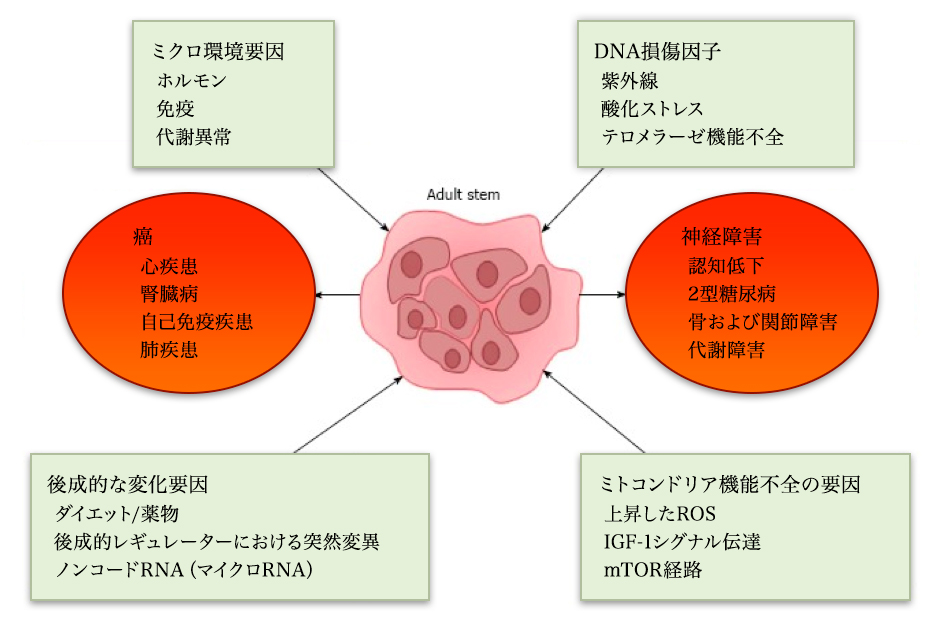
ABOUT STEMCELL

What are stem cells?

Omotesando Helene Clinic has acquired a plan number for providing second-class regenerative medicine.

Our clinic complies with the regulations of the Ministry of Health, Labor and Welfare's Regenerative Medicine Act, and has already received 15 regenerative medicine provision plans to date (as of June 2024), and has obtained an ISO9001 quality management system certification.

What is a stem cell? 

We all have the ability to regenerate and replenish lost cells in our bodies, like skin and blood, in order to maintain tissues that are short-lived and constantly replaced. have cells Cells with these abilities are called “stem cells”. To be called a stem cell, the following two abilities are essential. One is the ability to create various cells that make up our bodies, such as skin, red blood cells, and platelets (differentiation ability), and the other is the ability to divide into cells that have exactly the same ability as ourselves ( self-replicating).

Stem cells can be broadly divided into two types. One type is stem cells that continue to replace lost cells in specific tissues and organs, such as skin and blood. This type of stem cell is called "tissue stem cell." Tissue stem cells cannot become anything; they have a specific role, such as hematopoietic stem cells that produce blood, or neural stem cells that produce the nervous system. The other type is "pluripotent stem cell," which, like embryonic stem cells (ES cells), can produce any cell in our body. In other words, pluripotent stem cells can also produce various tissue stem cells that exist in our body. iPS cells (induced pluripotent stem cells) are "pluripotent stem cells" artificially created from ordinary cells.
These properties of stem cells are being utilized in research into new treatments known as "regenerative medicine," which uses cells themselves as medicine to treat injuries and diseases, and in research into the mechanisms of disease by recreating the state of internal cells outside the body.
Matters concerning the content and costs of treatments normally required for elective medical treatment

MSC PRINCIPLE

MSC principle

Effects of MSCs

Effect of MSC transplantation on diabetic cardiomyopathy. (A) MSC increases MMP-2 activation, suppresses activation of MMP-9, and attenuates cardiac remodeling. (B) MSCs produce VEGF, IGF-1, AM, HGF and stimulate myogenesis and angiogenesis in damaged myocardium. (C) Via differentiation into cardiomyocytes and vascular endothelial cells, MSC improves myocardial perfusion and myocardial regeneration. Abbreviations: AM, Adrenomedullin; HGF, Hepatocyte growth factor; IGF-1, Insulin-like growth factor-1; MMP, Matrix metalloprotease; MSC, Mesenchymal stem cell; VEGF, Vascular endothelial growth factor

Effects of MSC treatment on diabetic polyneuropathy

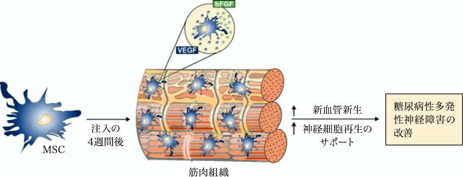
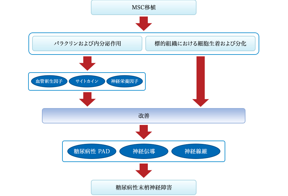
The effect of MSC treatment on diabetic polyneuropathy. Four weeks after intramuscular injection, MSCs deposit in the interstitial space of myofibers through the production of bFGF and VEGF, and induce neovascularization and support regeneration of neuronal cells leading to amelioration of diabetic polyneuropathy. Abbreviations: bFGF, Basic fibroblast growth factor; MSC, Mesenchymal stem cells; VEGF, Vascular endothelial growth factor

Image of mesenchymal stem cells

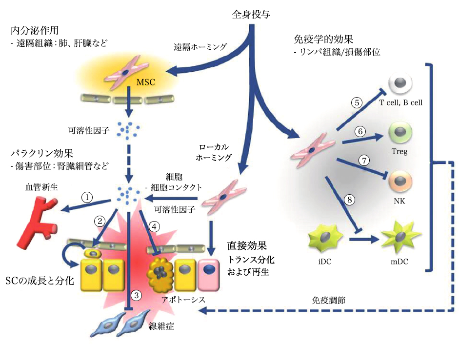
Systemic administration of mesenchymal stem cells can induce endocrine or local paracrine effects, including cell-mediated actions: 1) vascular endothelial growth factor (VEGF), insulin-like growth factor 1 (IGF-1), monocyte chemoattractant protein 1 (MCP1), basic fibroblast growth factor (bFGF), interleukin 6 (IL-6); 2) stem cell proliferation and differentiation: stem cell factor (SCF), leukemia inhibitory factor (LIF), macrophage colony-stimulating factor (MCSF), stromal cell-derived factor 1 (SDF1), angiopoietin 1, and activin A; 3) inhibition of fibrosis: hepatocyte growth factor (HGF), bFGF, and adrenomedullin (ADM); and 4) inhibition of apoptosis: VEGF, HGF, IGF-1, transforming growth factor (TGF)-β, bFGF, granulocyte-macrophage colony-stimulating factor (GM-CSF), activin A, and thrombospondin 1. Immune-mediated effects include (5-8): 5) Suppression of T and B cells: human leukocyte antigen G5 (HLA G5), HGF, inducible nitric oxide synthase (iNOS), indoleamine 2,3-dioxygenase (IDO), prostaglandin E2 (PGE2), bFGF, and TGFβ; 6) Induction of differentiation and proliferation of regulatory T cells (Tregs) through TGFβ expression; 7) Inhibition of natural killer (NK) cells through secretion of IDO, PGE2, and TGFβ; 8) Inhibition of dendritic cell (DC) maturation through secretion of PGE2.
Figure "Stem Cell Res Ther" reproduced by Carrión and Figueroa. 2011 May 11;2(3):23.

Diagram of NK cells

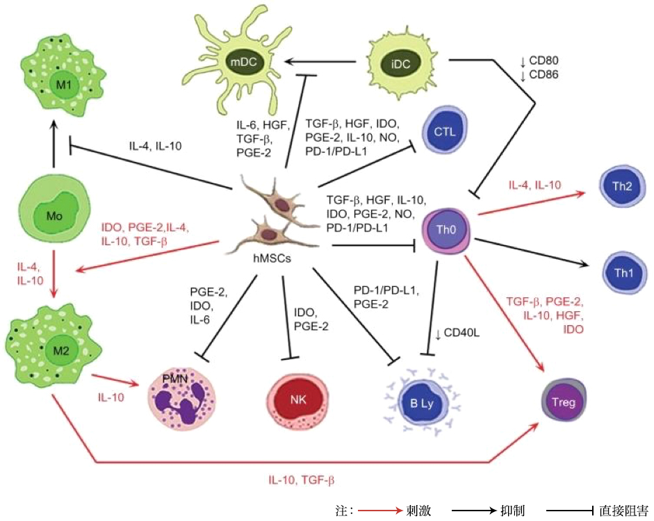
Note: Red arrow: stimulation; black arrow: inhibition; unhooked arrow: direct inhibition.
Abbreviations: iDC, immature dendritic cell; IL, interleukin; HGF, hepatocyte growth factor; TGF-β, transforming growth factor-β; PGE-2, prostaglandin E2; IDO, indoleamine 2,3-dioxygenase; NO, nitric oxide; PD-L1, programmed death ligand 1; hMSC, human mesenchymal stem cell; Treg, T regulatory; Th, T helper; CTL, cytotoxic T cell; mDC, mature dendritic cell; PD-1, programmed cell death protein 1; PMN, polymorphonuclear leukocyte; NK, NK cell

Local uptake in the anterior ventricular region of the animal heart

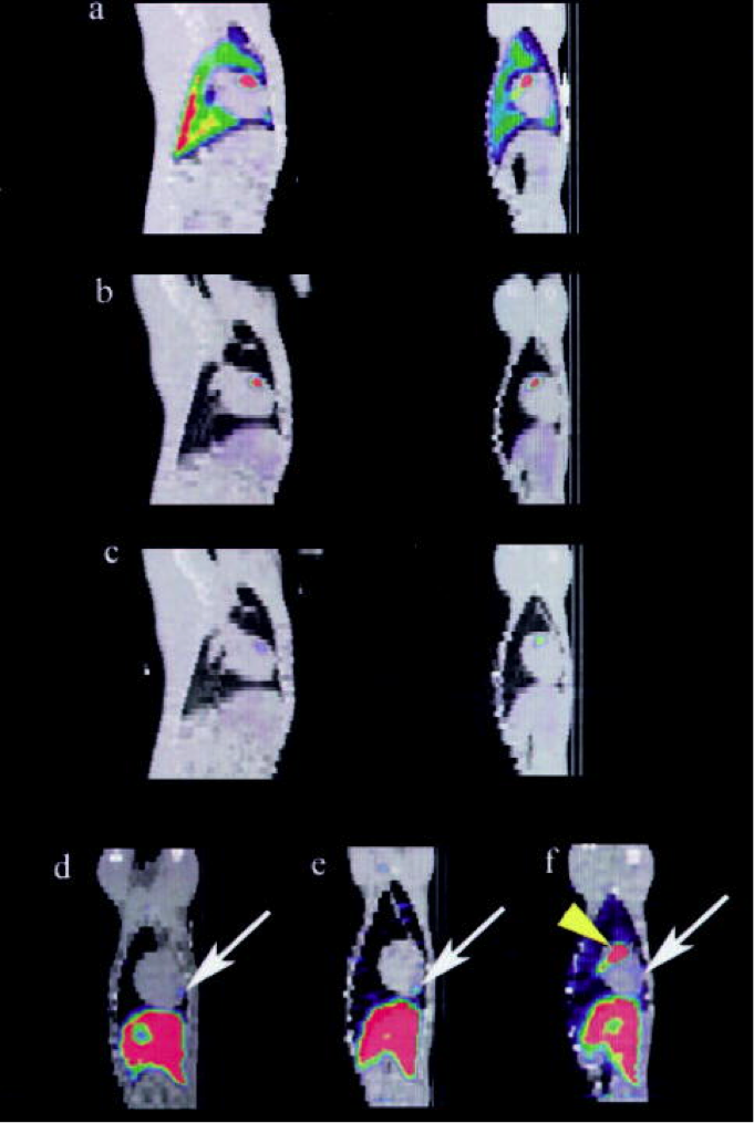
The sagittal plane (left) of the SPECT / CT image fused to day 1 (a), day 2 (b) and day 7 (c) showing local uptake in the anterior ventricular region of the animal’s heart, and this is the coronal plane (right). At the last imaging time (day 5 to 8), the anterior apical area of the MSC uptake (arrow) is shown in three representative animals in the coronal structure map. The vertex distribution ahead of this existed regardless. (Only yellow arrow of f)

isolated bone marrow stromal cells

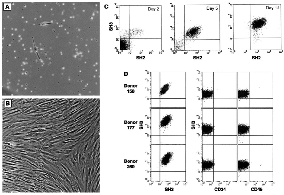
Characteristics of isolated bone marrow stromal cells. Cells are cultured from the bone marrow after density fractionation and are shown as (A) 48 hours after plating and (B) 10 days after plating. (C) Flow cytometry shows enrichment of these cultured cells. Results were obtained on day 2, day 5, and day 14 of culture using antibodies SH2 and SH3 generated against surface markers. (11) On day 14, the cells were 95-99% homogeneous and the reaction to the antigen CD14, CD34 (Becton-Dickinson), or CD45 (Pharmingen) common to hematopoietic cells was negative. (D) Homogeneity and reproducibility of the isolation procedure was demonstrated by flow cytometry

DISEASE

Disease

Clinical trial data
Indications used with MSCs in clinical trials. Data from 352 registered clinical trials
Indications Being Addressed using MSCs in Clinical Trials. Data for 352 registered clinical trials.
Citation// Stem Cell Therapies in Clinical Trials: Progress and Challenges. Trounson, Alan et al. Cell Stem Cell , Volume 17 , Issue 1 , 11 – 22

Disease · Unchanging

pluripotent stem cells
Pluripotent stem cells have remarkable self-renewal capabilities and can differentiate into multiple diverse cell types. Evidence is gradually mounting that the aging process can adversely affect stem cells. As stem cells senesce, their regenerative capacity declines and their ability to differentiate into various cell types changes. Therefore, it has been suggested that the decline in stem cell function due to aging may play an important role in the pathophysiology of various aging-associated diseases. Understanding the role of the aging process in stem cell function is important not only for understanding the pathophysiology of aging-associated diseases but also for developing effective stem cell-based therapies to treat aging-associated diseases in the future. This review article focuses on the fundamentals of stem cell dysfunction associated with various aging-associated diseases. We then discuss several concepts regarding the mechanisms that may be responsible for aging-associated stem cell dysfunction. We also briefly discuss current potential therapies for aging-associated stem cell deficiencies that are currently under development.
Citation// World J Exp Med. 2017 Feb 20; 7(1): 1–10.Effect of aging on stem cells. Abu Shufian Ishtiaq Ahmed,et al

Disease · Liver / Glycosuria

stem cells and liver

Regenerative medicine is transitioning to clinical programs using stem cell / progenitor cell therapy for repair of damaged organs. Liver and pancreas, organs that share the endodermal stem cell population, biliary stem cells located in the biliary trunk (hBTSC) are briefly described. They are precursors of hepatic stem / progenitor cells in the herring canal and are progenitor cells of the pancreatic duct. They produce a mature line along the radial axis within the bile duct wall and a proximal-distal axis starting at the duodenum and ending at mature cells in the liver or pancreas. Clinical trials have been conducted over the years to evaluate the effects of stem cells transplanted into the hepatic arteries of various liver disease patients (liver stem / progenitor cells from fetal liver). Immunosuppression was not necessary. All control subjects given the given criteria died within 1 year or the liver function declined. Subjects transplanted with 100-150 million liver stem / progenitor cells showed improvement in liver function and survival over several years. Evaluation of the safety and effectiveness of transplantation is still under development. Although stem cell therapy for diabetes using hBTSC is still being studied, it is likely to be done after ongoing preclinical studies. In addition mesenchymal stem cells (MSC) and hematopoietic stem cells (HSC) are used in patients with chronic liver disease or diabetes. MSCs are shown to be effective through paracrine of trophic factors and immune regulators and inefficient lineage restriction to mature parenchymal cells or pancreatic islet cells is limited. The effect of HSC is mainly due to regulation of the immune system.

Stem Cells. 2013 Oct;31(10):2047-60. doi: 10.1002/stem.1457. Concise review: clinical programs of stem cell therapies for liver and pancreas.Lanzoni G1, Oikawa T

Diabetes

QinanWu, Bing Chen, and Ziwen Liang,
Mesenchymal Stem Cells as a Prospective Therapy for the Diabetic Foot
Stem Cells International Volume 2016, Article ID 4612167, 18 pages http://dx.doi.org/10.1155/2016/4612167
Diabetic PAD

Figure 1: Mechanism of the effect of MSC transplantation on diabetic PAD. Mechanism of recovery effect mediated by stem cell transplantation from two pathways: One is the secretion of angiogenic factors and cytokines, and the other is transplantation and differentiation into tissue constituents of cells. Stem cells can specifically improve the local secretion and expression of angiogenic factors and cytokines, contribute to the reconstruction of the microcirculatory system and improvement of blood flow and islet β-cell function, leading to improvement of diabetic PAD. The stem cells can also differentiate into endothelial cells to achieve recovery of endothelial cell dysfunction. These effects may be related to miRNA and MEX.

diabetic wound

Figure 2: Mechanism of the effect of MSC transplantation on diabetic wounds. Diabetes wound recovery by MSC grafting from three pathways: First is angiogenesis and secretion of factors and cytokines, and second is regulation of the immune system, and third is transplantation and differentiation into tissue constituents of cells is there. Stem cells can specifically improve the local secretion and expression of angiogenic factors and cytokines that contribute to the improvement of diabetic PAD and diabetes. Also, stem cells can regulate the activity of T cells, natural killer cells, macrophages, and dendritic cells and can inhibit infection and inflammatory reactions. In addition, MSCs can differentiate into target tissues and achieve repair. These effects may be related to miRNA and MEX.

MSC transplantation

Figure 3: Mechanism of the effect of MSC transplantation on diabetic neuropathy. Mechanism of recovery effect mediated by stem cell transplantation arising from two pathways: One is the secretion of angiogenic factors, cytokines and neurotrophic factors, and the other is transplantation and differentiation into tissue constituents of cells. Stem cells can specifically improve the local secretion and expression of angiogenic factors and cytokines, contribute to the improvement of diabetic PAD and diabetes themselves, leading to amelioration of diabetic neuropathy. Neurotrophic factors can also improve nerve fiber dysfunction and nerve conduction. In addition, stem cells can differentiate into target tissues and achieve repair.

Kidney failure

Alfonso Eirin and Lilach O Lerman* Mesenchymal stem cell treatment for chronic renal failure,
Stem Cell Research & Therapy 2014, 5:83 http://stemcellres.com/content/5/4/83
Mesenchymal stem cell therapy

In animals undergoing mesenchymal stem cell therapy, stenosis – renal microvascular loss and fibrosis decreased. Above: Representative microcomputerized tomography of the kidney segment capturing the improved microvascular structure in atherosclerotic renal artery stenosis, pigs who underwent percutaneous transluminal renal angioplasty (PTRA) three dimensional In the image, adrenal infusion of adipose tissue-derived mesenchymal stem cells (MSC) was performed 4 weeks earlier. Bottom: Representative kidney trichrome staining showing a decrease in MSAS fibrosis of ARAS + PTRA + pig (× 40, blue)

Clinical application example of MSC: diabetes

Stem cell transplantation can be a safe and effective treatment for patients with DM. In this series of trials, the best treatment outcome was achieved with D34 + HSC therapy for T1DM, but the worst results with HUCB for T1DM were observed. Diabetic ketoacidosis interferes with the therapeutic effect.

Examples of clinical applications of MSC

A line graph showing the change in C-peptide and HbA1c levels at baseline, 3 months, 6 months, and 12 months after stem cell treatment in T1DM patients. All data are expressed as mean ± SEM. **** P 0.0001

The outcome for stem cell therapy for T2DM

Stem cell therapy for type 2 DM.

Bar graph showing changes in C-peptide and HbA1c levels

A-D)A bar graph showing the baseline after administration of different types of stem cells, as well as the changes in C – peptide and HbA 1c levels after 12 months. (EF) T2D injected UCB and BM-MNC into the pancreas (n = 3 and n = 107) while intravenously injected with UC-MSC and PD-MSC (n = 22 and n = 10 respectively) A line graph showing the change in C-peptide and HbA1c levels at baseline, 3 months, 6 months, and 12 months after treatment.

Citation// PLoS One. 2016 Apr 13;11(4):e0151938. Clinical Efficacy of Stem Cell Therapy for Diabetes Mellitus: A Meta-Analysis. El-Badawy A, El-Badri N.

Disease · Hair follicle

Nat Commun. 2012 Apr 17;3:784. doi: 10.1038/ncomms1784.
Fully functional hair follicle regeneration through the rearrangement of stem cells and their niches.
Toyoshima KE1, Asakawa K, Ishibashi N, Toki H, Ogawa M, Hasegawa T, Irié T, Tachikawa T, Sato A, Takeda A, Tsuji T.

Overview
Organ replacement regenerative medicine promises to enable the replacement of organs damaged by disease, injury, or aging in the foreseeable future. Here, we demonstrate fully functional organ regeneration via intradermal transplantation of bioengineered bone and blastoderm. The blastoderm and ovule are reconstituted with embryonic skin-derived cells and adult stem cell field-derived cells, respectively. The bioengineered hair follicles develop the correct architecture and form appropriate connections with surrounding host tissues, such as the epidermis, hindlimb muscle, and nerve fibers. The bioengineered hair follicles also exhibit restored hair cycle and hair head formation through reorganization of hair follicle stem cells and their niches. This study therefore reveals the potential of adult tissue-derived hair follicle stem cells as a bioengineered organ replacement therapy.

Organ replacement regenerative medicine

(a) Schematic of the method used for making and transplanting bioengineered hair follicle embryos. (b) Phase difference images of dorsal skin, tissues, dissociated single cells and bioengineered hair follicle embryos of mouse embryos reconstructed using the organ germ method using nylon thread (arrow head). Scale, 200 μm. (c) Histological analysis of isolated tentacles from adult mice. Macroscopic and H & E staining bristles are shown on the left two panels. The dashed line (red) by macro morphological observation (left) and H & E staining (right) shows the interface of bulge and SB region. The box area on the left panel is stained with H & E to show the bulge and the SB area is shown on the higher panel in the higher panel. The bulge region was immunostained with anti-CD49f (red, left) and anti-CD34 (red, middle) antibody and Hoechst 33258 dye (blue). The black dashed line at high magnification H & E indicates the interface of the epithelium of the follicle. IF, funnel; RW, annular body; half of hair follicle. Scale, 100 μm. (d) Histological and ALP analysis of initial regeneration of tactile valve area and DP cells. Hair bulbs (2 left) and cultured DP cells (2 right) were analyzed by ALP enzyme staining. The red dotted line shows Auber’s line. Scale “, 100 μm. (e) Longitudinal section of bioengineered hair during the eruption and growth process, mediated by the inter-epithelial tissue-interlocking plastic device (guided). Corresponding items are (without guide) skin It is shown as cystogenesis with bioengineered hair follicles 14 days after transplantation H & E staining (top) of bioengineered hair follicles on days 0, 3 and 14 post- Fracture microscope (bottom) Scale, 100 μm (f) Healing of wounds immediately after implantation (left), day 3 (center) on day 0, ejection of hair shafts on days 14 and 37 , And macrostructural observation of grown hair during development and growth of bioengineered chest (upper) and spleen (lower), including growth Scale, 1.0 mm.

Analysis sample

(a) Histological and immunohistochemical analysis of bioengineered hair (upper) and tentacular (central) vesicles. The box area of the low magnification H & E panel is shown at higher magnification in the right panel. Arrows indicate sebaceous glands. Scale, 100μm. Bioengineered follicle hair bulbs were immunostained with anti-versican (lower left) and α-SMA (arrow head, lower right) antibodies and also enzymatically stained for ALP. (Lower center) scale, 50 μm. (b) Bioengineered human hair produced by transplantation of biologically treated hair follicle embryos were reconstituted by intact DP of bulge derived epithelial cells and human scalp follicles. Human hair by bioengineering was photographed (microscopic examination) on day 21 after transplantation and analyzed by H & E staining. According to nuclear morphological characteristics (right panel), species identification of bioengineered hair follicles was analyzed. The enclosed area of the inset is high magnification. Scale, microscope 500 μm, H & E 100 μm, nuclear dyeing 20 μm. (c) Dense intradermal transplantation of bioengineered follicular pathogens. A total of 28 independent bioengineered hair follicle germs were transplanted into the uterine cervical skin of the mouse and showed high density hair growth on day 21 after transplantation. Scale, 5 mm.

Results of regeneration of the bulge region

Bioengineered hairs and tentacles were connected with other tissues such as nerve fibers, retractor muscles and striatum muscles derived from host or donor cells. The bioengineered hair bound smooth muscle as a result of regeneration of the NPNT-expressing bulge region, similar to the natural mass. Neither NPNT expression nor smooth muscle binding was detected in the bioengineered hair bulge region.

Quote/Fully functional hair follicle regeneration through the rearrangement of stem cells and their niches. Koh-ei Toyoshima, Kyosuke Asakawa, Naoko Ishibashi, Hiroshi Toki, Miho Ogawa, Tomoko Hasegawa, Tarou Irié, Tetsuhiko Tachikawa, Akio Sato, Akira Takeda & Takashi Tsuji. Nature Communications 3, Article number: 784 (2012)
doi:10.1038/ncomms1784

Other diseases · Parkinson

Schematic representation of stem cell induction, differentiation and application

Schematic of induction, differentiation and application of stem cells currently available for PD research and therapy. The above stem cells can be divided into four categories: ESC, NSC, MSC, and iPSC, with gradually decreasing differentiation totipotency. (1) ESC originating mainly from the blastocyst inside the blastocyst can simultaneously differentiate into endoderm, mesoderm and ectoderm under normal circumstances. In some cases ESC can also be induced to differentiate into NSC and MSC. (2) NSC isolated directly from specific brain niche or reprogrammed from fibroblasts can perform neural lineage differentiation into neurons and almost all glial cells. (3) MSC is mainly derived from mesenchymal tissue and can differentiate into almost all cells of mesoderm origin. Remarkably, MSC can be induced to differentiate into DA neurons even under certain combinations of induction protocols. (4) iPSCs that can be re-differentiated from adult human somatic cells (such as fibroblasts) by introducing OSKM (Oct 3/4, Sox 2, Klf 4, and c-Myc) It is a promising source of stem cells to have. Based on the GMP standard, the stem cells and terminally differentiated cells described above can be further sorted, purified and expanded for application to disease model, drug screening, and CRT practice. For example, ESC, MSC, NSC, and DA neurons are used below. (I) Preparation of PD model (ii) Potential drug screening; (iii) CRT treatment of PD Front. Aging Neurosci., 31 May 2016. A Compendium of Preparation and Application of Stem Cells in Parkinson’s Disease: Current Status and Future Prospects. Yan Shen, Jinsha Huang

RISK

RISK

Clinical trial risks

Background:
Mesenchymal stromal cells (MSCs, "adult stem cells") are widely used experimentally in a variety of clinical settings. There is interest in using these cells in serious illnesses, but the safety profile of these cells is not well known. We conducted a systematic review of clinical trials investigating the use of MSCs to evaluate their safety.

Methods and Results:
MEDLINE, EMBASE, and CENTRAL (up to June 2011) were searched. Clinical trials were conducted using intravascular delivery (intravenous or intra-arterial) of MSCs in adults or mixed adult and pediatric groups. Studies using differentiated MSCs or additional cell types were excluded. Primary endpoints were classified according to immediate events (acute infusion toxicity, fever), organ system complications, infection, and longer-term adverse events (death, malignancy). 2347 citations were reviewed, and 36 studies met the inclusion criteria. The 1012 participants included patients being treated for ischemic stroke, Crohn's disease, cardiomyopathy, myocardial infarction, graft-versus-host disease, and healthy volunteers. Eight studies were randomized controlled trials (RCTs) with 321 participants.

Multilingual summary:
Multilingual summary text

Citation// PLoS One. 2012;7(10):e47559. doi: 10.1371/journal.pone.0047559. Epub 2012 Oct 25.
Safety of cell therapy with mesenchymal stromal cells (SafeCell): a systematic review and meta-analysis of clinical trials. Lalu MM, McIntyre L, Pugliese C, Fergusson D, Winston BW, Marshall JC, Granton J, Stewart DJ; Canadian Critical Care Trials Group.

×
×
×
×
×
×
×
×
×
×
×
×

×

×
×

×
×
×
×
×

×
×
×
×
culture video
×
×
Former Health, Labor and Welfare Minister Ritsuo Hosokawa
×

×

×
×
×
×
×

×

×

×

×
×